Home » Without Label » Back Of Neck Anatomy : Posterior Neck Anatomy(4) - The cervical spine supports the weight and movement of your head and protects the nerves exiting your brain.
Back Of Neck Anatomy : Posterior Neck Anatomy(4) - The cervical spine supports the weight and movement of your head and protects the nerves exiting your brain.
Back Of Neck Anatomy : Posterior Neck Anatomy(4) - The cervical spine supports the weight and movement of your head and protects the nerves exiting your brain.. One of the functions of the neck is to act as a conduit for nerves and vessels. Anatomy of back of human neck, anatomy of the back and neck, anatomy of the back of the neck, anatomy of the back of the neck muscles, anatomy of the back of your. The anterior, and the posterior, triangles of the neck. Neck anatomy explained the neck begins at the base of the skull and connects to the thoracic spine (the upper back). The nerves of the head and neck include the most vital and important organs of the nervous system — the brain and spinal cord — as well as the organs of the special senses.
The back of the neck is called the posterior cervical area. However, other muscles also constitute the neck. It is more prominent in men. The anterior triangle of the neck is made by the anterior border of the sternocleidomastoid muscle, the inferior border of the mandible and the midline of the neck. Back pain is common and might be caused by a problem with a muscle.
Neck muscles of the anterior surface. from anatomyatlases.org The back of the neck is called the posterior cervical area. Anatomy of back of human neck, anatomy of the back and neck, anatomy of the back of the neck, anatomy of the back of the neck muscles, anatomy of the back of your. The neck is a complex anatomic region between the head and the body. In the front, the neck extends from the bottom part of the mandible (lower jaw bone) to the bones of the upper chest and shoulders (including the sternum and collar bones). The neck is connected to the upper back through a series of seven vertebral segments. This guide gives a general overview of the anatomy of the neck. Hyoid bone explore study unit There are two main triangles;
The back of the neck is mostly comprised of muscles, as well as the spine.
Neck anatomy explained the neck begins at the base of the skull and connects to the thoracic spine (the upper back). The suprahyoid and infrahyoid muscles. Anatomy of back of human neck, anatomy of the back and neck, anatomy of the back of the neck, anatomy of the back of the neck muscles, anatomy of the back of your. The laryngeal prominence, more commonly known as the adam's apple, is a noticeable external neck feature. The cervical spine protects the nerves connecting to the brain, allowing the head to move freely while supporting its. These muscles give the sides of the neck their. The neck is the part of the body on many vertebrates that connects the head with the torso and provides the mobility and movements of the head. Cervical spine anatomy video the cervical spine has 7 stacked bones called vertebrae, labeled c1 through c7. It runs down the back part of the neck, and opens into the external jugular vein just below the middle of its. The cervical spine supports the weight and movement of your head and protects the nerves exiting your brain. There are numerous muscles associated with the throat, the hyoid bone and the vertebral column; Neck muscles can be strained from poor posture — whether it's leaning over your computer or hunching over your workbench. The cervical spine, your neck, is a complex structure making up the first region of the spinal column starting immediately below the skull and ending at the first thoracic vertebra.
They start at the top of the neck and go down to the tailbone. The anterior triangle of the neck is made by the anterior border of the sternocleidomastoid muscle, the inferior border of the mandible and the midline of the neck. It runs from the neck to the upper back. Cervical spine anatomy video the cervical spine has 7 stacked bones called vertebrae, labeled c1 through c7. Within these compartments, the neck houses the cervical vertebrae and cervical part of the spinal cord, upper parts of.
Posterior Neck Anatomy(5) from www.medicalexhibits.com Cervical spine anatomy video the cervical spine has 7 stacked bones called vertebrae, labeled c1 through c7. It runs from the neck to the upper back. The posterior external jugular vein (v. The suprahyoid and infrahyoid muscles. There are two main triangles; One of the functions of the neck is to act as a conduit for nerves and vessels. In addition, in this region we also find the major cranial and spinal nerves that connect the central nervous system to the organs, skin, and muscles of the head and neck. Back pain is common and might be caused by a problem with a muscle.
See human neck anatomy stock video clips.
The cervical spine, your neck, is a complex structure making up the first region of the spinal column starting immediately below the skull and ending at the first thoracic vertebra. The spinal cord is a bundle of nerves that carry electrical signals between the brain and the rest of the body. Think of it like a jigsaw puzzle, all the pieces fit in together and are required to get the full picture as to how it works. Neck anatomy nerves picture there are 8 spinal nerves that originate from the cervical spine. The anterior triangle of the neck is made by the anterior border of the sternocleidomastoid muscle, the inferior border of the mandible and the midline of the neck. In addition, in this region we also find the major cranial and spinal nerves that connect the central nervous system to the organs, skin, and muscles of the head and neck. It is made up of bones, discs, muscles, ligaments, nerves and tendons. The muscles of the neck run from the base of the skull to the upper back and work together to bend the head and assist in breathing. It is more prominent in men. One of the most important jobs of the cervical spine is to protect the spinal cord as it travels through the neck to innervate the rest of the body. It runs down the back part of the neck, and opens into the external jugular vein just below the middle of its. The back of the neck is called the posterior cervical area. It runs from the back of the head to the small of the back.
It consists of seven vertebrae. In the front, the neck extends from the bottom part of the mandible (lower jaw bone) to the bones of the upper chest and shoulders (including the sternum and collar bones). Browse 3,107 anatomy of neck and shoulder stock photos and images available, or start a new search to explore more stock photos and images. Spinal cord anatomy in the neck. Back pain is common and might be caused by a problem with a muscle.
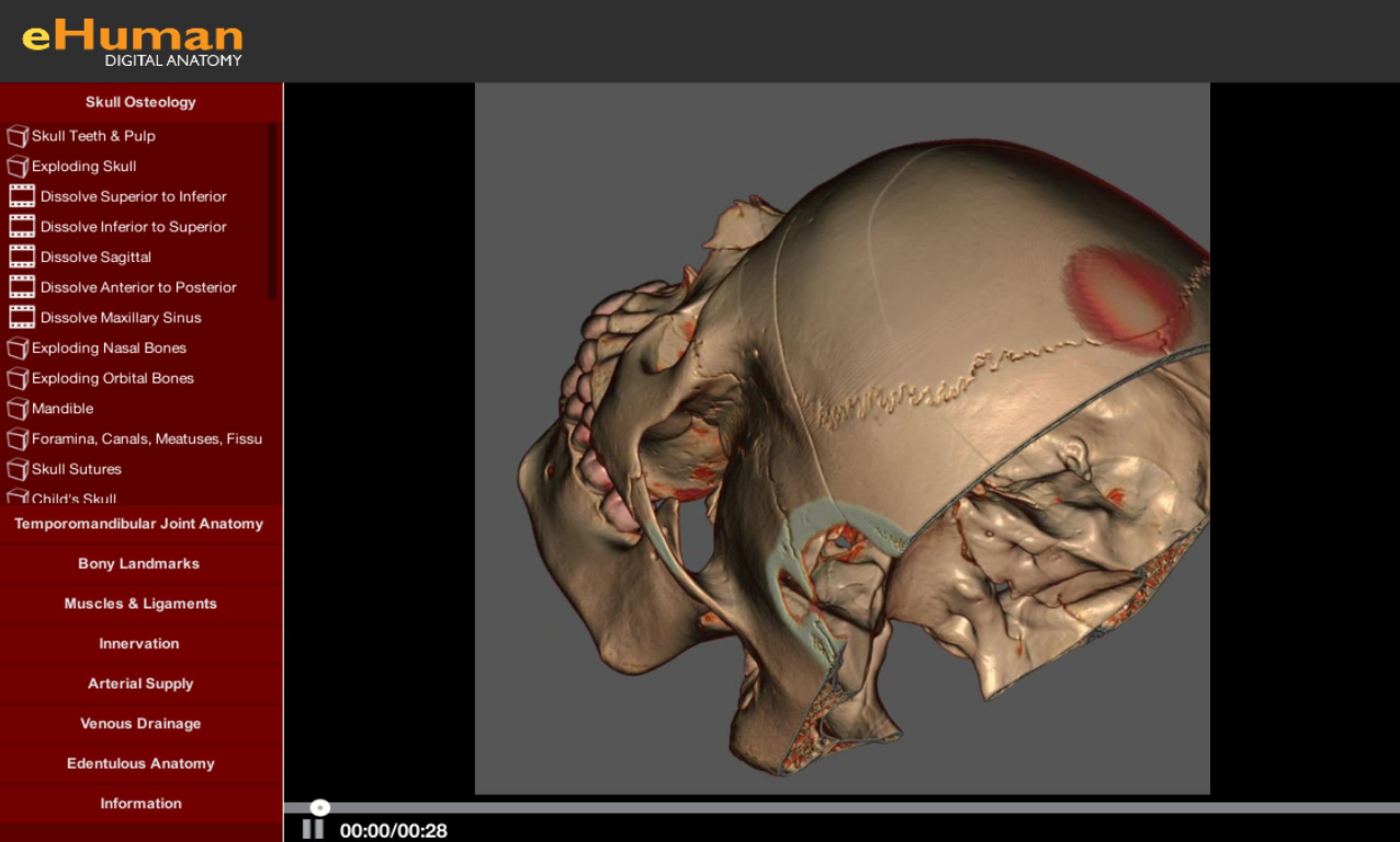
Head and Neck Anatomy | eHuman from ehuman.com An area called the occiput. The top of the cervical spine connects to the skull, and the bottom connects to the upper back at about shoulder level. Neck muscles can be strained from poor posture — whether it's leaning over your computer or hunching over your workbench. The occipital bone is the only bone in your head that connects with your cervical spine (neck). Spinal cord anatomy in the neck. It runs from the neck to the upper back. The anterior, and the posterior, triangles of the neck. Neck anatomy nerves picture there are 8 spinal nerves that originate from the cervical spine.
Each nerve provides sensation to a specific area of the body called a dermatome.
There are two main triangles; The cervical spine protects the nerves connecting to the brain, allowing the head to move freely while supporting its. The neck is a complex anatomic region between the head and the body. In addition to reading this article, be sure to watch our cervical spine anatomy animated tutorial video. Back pain is common and might be caused by a problem with a muscle. See anatomy of the head and neck stock video clips. The anterior triangle of the neck is made by the anterior border of the sternocleidomastoid muscle, the inferior border of the mandible and the midline of the neck. These muscles give the sides of the neck their. The occipital bone is the only bone in your head that connects with your cervical spine (neck). It runs down the back part of the neck, and opens into the external jugular vein just below the middle of its. They start at the top of the neck and go down to the tailbone. In addition, in this region we also find the major cranial and spinal nerves that connect the central nervous system to the organs, skin, and muscles of the head and neck. Muscle head anatomy vocal organ diagram female neck anatomy neck wireframe head neck human anatomy head artery anatomy face pharynx vector neck degree head anatomy 3d.